Speeding up cancer diagnosis during surgery
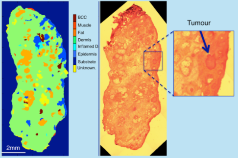
The availability of diagnosis and tumour imaging during surgery is extremely valuable to surgeons who can excise and evaluate sequential layers of tissue to ensure the complete removal of tumour cells, while sparing as much healthy tissue as possible.
This surgical technique, called tissue-conserving surgery, has been increasingly employed in the treatment of cancer, such as skin or breast. However, ensuring the complete excision of the tumour remains one of the key challenges in tissue-conserving surgery. Failure to remove all tumour cells increases the risk of tumour recurrence and the need for secondary surgery.
Using optical microscopy to provide a molecular analysis of the tissue, we can produce images of individual cells and tumours with micron-scale spatial resolutions in only 20 minutes, making it clinically relevant to be used during surgery.
We are currently developing 2 devices for tumour detection: for skin cancer and breast cancer. Our next steps are to test the devices in 5 hospitals.