Biophotonic sensing and imaging
Using new optical and spectroscopic techniques we are developing new capabilities for studying biological materials at a nano- and micro-scale, enabling us to look into different regions of individual cells and enable imaging of biological materials with unprecedented informational detail.
Raman spectroscopy sensing
Biophotonics techniques based on Raman spectroscopy are being used to study nanomaterials and live cells and can detect molecular changes in tissues associated with either the cause or the effect of disease processes.
Advances and applications
A key advantage of Raman spectroscopy is that the information is obtained non-invasively and label-free, without need to damage cells or disturb their biological activity by using fluorescent labels or other contrast enhancing chemicals.
Raman sensing is being combined with novel nanomaterials developed at Nottingham to produce surface enhanced (SERS) biosensors capable of very low detection limits of biomarkers, integrated within microfluidic devices.
Current applications being explored include
- Understanding the interaction of cells with drugs toxicity assessment
- Differentiation of stem cells
- One-step capture and analysis of biomarkers in diagnostics
Experts:
Professor Ioan Notingher
Nottingham Nanoscience Group
Dr Kevin Webb
Optics and Photonics Research Group
Biophotonic imaging
Our recent developments in laser and light detection technology allow imaging of biological materials with unprecedented informational detail.
Advances and applications
We can now detect small chemical alterations in cells and tissue with sub-micron scale resolution, providing insight into dynamic processes and allowing label-free diagnosis of diseases.
Our strengths include label-free non-invasive methods, allowing rapid imaging of large areas, and following dynamic processes at a range of spatial scales including non-invasive imaging of molecular transport across tissue barriers.
We have developed novel methods in super-resolution microscopy and photoacoustic imaging as well as new methods for inexpensive and simple optical microscopy.
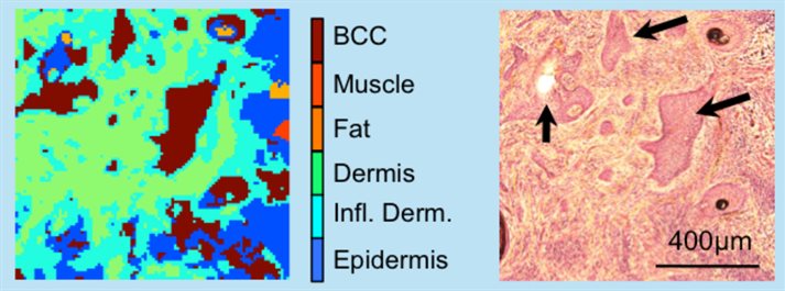
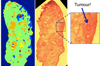
We are exploring applications in the imaging and diagnosis of cancer:
- Development of selective sampling methods for fast imaging of tissue.
- Spectral histopathology of skin and breast cancers.
Press release:
Speeding up cancer diagnosis during surgery
Experts:
Professor Ioan Notingher