Opto-electronic sensing
Opto-electric sensing in medical devices can measure a wide range of clinical parameters, and we have a strong track-record of translating our optical sensing devices into clinical use, through our excellent clinical collaborations at Nottingham University Hospitals NHS Trust and with industrial partners.
Optical fibre sensing
At just the width of a hair, cheap to purchase and highly flexible, optical fibres are ideal for incorporating into medical devices to provide enhanced function.
We have successfully incorporated our optical sensors in the development of critical care monitoring devices, and novel surgical devices
Measurement capabilities
Adding a functional chemical coating to the sensors has extended the range of parameters that can be measured to gases, biomarkers and drugs, and by embedding these functional sensors into textiles, we have opened up new clinical applications.
Our optical fibre sensors can measure
- Heartrate
- Oxygen saturation
- Mechanical properties eg absolute pressure
We are currently developing optical fibre chemical sensors, functionalised with nano-assembled thin film coatings. These can be highly sensitive and selective and can measure gases, biomarkers and drugs eg:
- Humidity
- pH
- Pressure
- Gases (eg Ammonia, Benzene, Chloroform, Toluene, Carbon dioxide, Carbon monoxide, Formaldehyde, Nitrogen dioxide)
- Biomarkers and drugs (eg Vancomycin, Streptavidin, Immunoglobulin)
For smart devices, contact Professor Steve Morgan
For functional sensing, contact Dr Serhiy Korposh
Smart textiles
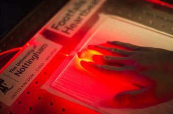
Through our collaboration with Footfalls and Heartbeats, we are developing textiles incorporating sensors. The revolutionary manufacturing process developed by Footfalls and Heartbeats creates a smart fabric which uses nano-scale interactions within the textile to make the fabric itself the sensor, avoiding the need for wires or miniature electronics.
Applications
Applications we are exploring include:
- Smart wound care – we can determine how tight a bandage is, and what are the levels of moisture within a wound dressing
- Diabetic foot care –using smart socks which monitor the blood flow in feet for diabetic patients, we can predict tissue breakdown in those with diabetes to help prevent diabetic foot ulcers
- Bedsores – by monitoring absolute pressure, we could predict and prevent bedsores
- Gait analysis – preventing falls.
For textile-based sensing contact Professor Steve Morgan
Optical endoscope
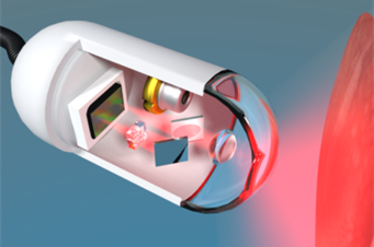
Optical endoscopes are being developed that improve detection of diseases such as cancer. Cancers that occur deep in the body are difficult to detect and treat, so these endoscopes, the size of a needle, will address this medical challenge.
Shrinking the microscope
Led by Dr George Gordon, a prototype holographic fibre endoscope has been designed and constructed, that images optical intensity, phase and polarisation.
This improves early detection of oesophageal cancer compared to conventional intensity-only endoscopes. The long (2m) and thin (500um diameter) geometry promises easier integration with existing clinical procedures, and paves the way for future imaging of previously inaccessible areas, e.g. the brain.
Low-cost imaging
Dr Gordon is exploring low-cost imaging to develop new capsule endoscopes for early cancer detection in the oesophagus and plasmonic metasurfaces for low-cost multispectral imaging.
Cancers that occur deep within the body, such as ovarian and pancreatic cancers, are difficult to detect and treat due to their inaccessibility via natural orifices. This typically makes them more deadly.
Dr Gordon’s research will address this problem by shrinking state-of-the-art microscopes to the size of a hair so they can fit inside a needle and take images deep inside the body, creating ultra-thin endoscopes. These will use nanotechnology to recreate the components of an optical microscope on the tip of these hair-thin fibres.
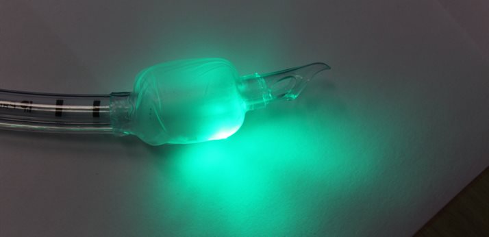
Fibre-optic ultrasonic imaging probe
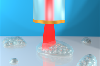
An ultrasonic imaging system, is being developed, which can be deployed on the tip of a hair-thin optical fibre, and will be insertable into the human body to visualise cell abnormalities in 3D.
World first probe set to examine hard-to-reach parts of the body
Led by Dr Salvatore La Cavera III, the new technology produces microscopic and nanoscopic resolution images that will one day help clinicians to examine cells inhabiting hard-to-reach parts of the body, such as the gastrointestinal tract, and offer more effective diagnoses for diseases ranging from gastric cancer to bacterial meningitis.
The EPSRC-funded innovation reduces the need for fluorescent labels, which can be harmful to human cells; and it its compact size has the potential to bring high performance imaging into clinical settings to improve patient care.
The findings are reported in Phonon imaging in 3D with a fibre probe (Nature journal, Light: Science & Applications)
Case studies
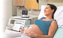
Monica Healthcare
A wireless device to overcome heart rate confusion between mother and unborn child during labour
Monica case study
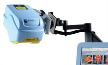
Moor Instruments
A new Laser Doppler Imaging device enabling clinicians to make more accurate decisions on the treatment of burns
Moor Instruments case study
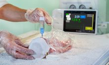
Surepulse Medical
A novel hands-free electronic heart rate monitor allowing continual monitoring of a baby’s heart-rate during resuscitation
SurePulse case study
Experts